Showing posts with label Anatomy. Show all posts

Saturday, November 19, 2011

BRS Gross Anatomy, 7th ed. 2012


This is a new edition of an established human gross anatomy review book that covers the material taught in courses in a concise and well organized manner. It provides brief explanations of gross anatomy concepts and terms and organizes muscles, blood vessels, and nerves in helpful tables. ..Unique features include the outline format that allows readers to quickly review without being distracted by unnecessary detail; the tests at the end of each chapter and the comprehensive test at the end of the book in a format similar to the USMLE exam...This is a helpful tool for students preparing for gross anatomy exams. It also can function as a quick reference for healthcare practitioners.

Sunday, October 2, 2011

Principles of Anatomy and Physiology 13th ed. 2012


The thirteenth edition of the phenomenally successful Principles of Anatomy and Physiology continues to set the standard for the discipline. The authors maintained a superb balance between structure and function and continue to emphasize the correlations between normal physiology and pathophysiology, normal anatomy and pathology, and homeostasis and homeostatic imbalances. The acclaimed illustration program continues to be refined and is unsurpassed in the market. The thirteenth edition is fully integrated with a host of innovative electronic media, including WileyPlus 5.0 (access purchased separately.) No other text and package offers a teaching and learning environment as rich and complete.


dOWNLOAd .

Saturday, October 1, 2011

Langman's Medical Embryology, 11th ed. 2009


Widely acclaimed for its clinical relevance and clear writing style, Langman's Medical Embryology provides a user-friendly and innovative approach to learning embryology. The revised eleventh edition presents medical and health professions students with a concise yet thorough description of embryology and its clinical significance, an awareness of which is essential in the diagnosis, and prevention of birth defects. It features outstanding full-color clinical images and photographs, end-of-chapter Summaries, Problems to Solve, and Clinical Correlates boxes. A companion Website includes fully searchable text, interactive review questions and problems, updated Simbryo animations of embryologic system development, and PowerPoint slides for instructors.


 dOWNLOAd . 

Monday, September 19, 2011

Netter's Clinical Anatomy, 2nd ed. 2010


Netter's Clinical Anatomy, 2nd Edition, by John T. Hansen, PhD, is an anatomy resource that clearly provides a comprehensive understanding of complex clinical anatomical concepts. Nearly 600 beautifully colored illustrations offer essential depictions of normal and pathologic anatomy and embryology, accompanied by supportive text to help you understand their clinical relevance. Short-answer review questions at the end of each chapter help you gauge your mastery of the material, and convenient online access equips you with the complete contents of the book, as well as additional illustrations and study questions. The result is a thorough but quick introduction to basic and clinical anatomy as well as a fast review source. Also available: Netter Atlas of Human Anatomy and Netter's Anatomy Flash Cards.
  • Presents nearly 600 beautifully colored illustrations which provide essential depictions of anatomy, embryology, and pathology to help you understand their clinical relevance.
  • Features Clinical Focus boxes throughout that present hundreds of well-illustrated clinical correlations which bridge anatomy to pathophysiology, and Feature and Characteristics boxes that explain the relation between structure and function.
  • Includes Muscle/Ligament/Joint Tables that summarize attachment points, actions, and other key information related to each structure, providing a quick overview of the MSK system
Download

Anatomy and Physiology: The Unity of Form and Function, 5th ed. 2009


Memorable stories must be effective in multiple ways. The story must paint a strong visual picture. The story must weave in tools to make the reader remember important events and understand their impact. Ken Saladin weaves graceful descriptions of human anatomy and physiology processes together with carefully selected clinical applications and fascinating stories from the history of medicine and evolutionary medicine to create a multi-layered story about the human body. A consistent set of chapter learning tools helps students identify and retain key concepts while the stunning visual program provides a realistic view of body structures and processes. The fifth edition is further improved by a complete integration of the text with extensive digital teaching and learning tools.


Thursday, April 14, 2011

Foundations of Manual Lymph Drainage, 3rd ed.


Chapters are structured according to anatomic regions, focusing on the lymphatic knots and their tributary regions in.


Surgical Anatomy of the Face, 2nd ed.


Atlas presents relevant facial anatomy from a surgical point of view. Includes such topics as facial contour analysis, facial embryogenesis, tissue foundation, anatomic regions, and variations in facial anatomy with race, sex, and age. High-quality drawing and photographs.


Human Dissection For Students Of Anatomy


Elearning video for students of anatomy.

FILE: 01 – parts 01-08.avi
01 – 08m00s – SKIN AND SUBCUTANEOUS TISSUE OF TRUNK AND UPPER THIGH
02 – 14m18s – INTRINSIC MUSCLES OF THE BACK / VERTEBRAL COLUMN / SPINAL MENINGES / SPINAL CORD
03 – 10m31s – THE ANTERIOR BODY WALL
04 – 26m43s – LUNGS PLEURAL SACS / HEART PERICARDIAL SACS
05 – 21m09s – POSTERIOR THORACIC WALL
06 – 24m49s – PERITONEAL CAVITY
07 – 23m49s – ABDOMINAL ORGANS
08 – 10m09s – ABDOMINAL AUTONOMICS.

FILE: 02 – parts 09-15.avi
09 – 14m51s – SPLIT PELVIS
10 – 16m30s – THE PELVIC OUTLET / PERINEUM
11 – 12m03s – SUPERFICIAL FACE
12 – 12m25s – INFRATEMPORAL FOSSA
13 – 14m23s – SUPERFICIAL NECK
14 – 15m12s – DEEP NECK
15 – 25m55s – CRANIAL CONTENTS

FILE: 03 – parts 16-21.avi
16 – 11m21s – THE ORBIT
17 – 05m28s – THE EAR
18 – 34m45s – CERVICAL JOINTS, MOUTH PHARYNX
19 – 10m43s – NASAL CAVITY SINUSES
20 – 06m56s – SUPERFICIAL DISSECTION OF THE LIMBS
21 – 12m15s – AXILLARY FOSSA

FILE: 04 – parts 22-27.avi
22 – 17m19s – SHOULDER AND ARM MUSCLES
23 – 20m18s – FOREARM AND HAND
24 – 07m40s – JOINTS OF THE UPPER EXTREMITY
25 – 14m38s – MUSCLES, VESSELS, AND NERVES OF THE HIP AND THIGH
26 – 14m07s – LEG AND FOOT
27 – 09m09s – JOINTS OF THE LOWER EXTREMITY


Download links:part-1 
part-2 
part-3 
part-4 
part-5 
part-6 
part-7 
part-8 
part-9 
part-10 

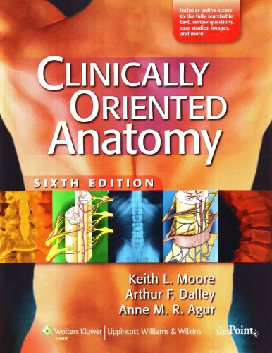
Clinically Oriented Anatomy, 6th ed.


Clinically Oriented Anatomy, Sixth Edition provides first-year medical and allied health students with the clinically oriented anatomical information that they need in study and practice. This best-selling textbook is renowned for its comprehensive coverage of anatomy, presented as it relates to the practice of medicine, dentistry, and physical therapy. The Sixth Edition features a modified interior design with new and improved artwork that further enhances the user-friendliness of the text. The clinical Blue Boxes are now grouped to reduce interruption of text and are categorized with icons to promote easier comprehension of clinical information. The Bottom Line summaries have been refined to clearly call out key points for quick study. A companion Website offers the fully searchable text, interactive USMLE-style questions, and video animations. Online faculty resources include an Image Bank, Test Generator, and Course Outlines.


Grant's Atlas of Anatomy, 12th ed.


A cornerstone of gross anatomy since 1943, Grant's Atlas of Anatomy continues to reach students worldwide with its realistic illustrations, detailed surface anatomy photos, clinical relevance, and muscle tables. Renowned for its accuracy, pedagogy, and clinical relevance, the new edition of this classic anatomy atlas features many improvements, including updated artwork, more vital tissue colors, new conceptual diagrams, vibrantly re-colored illustrations, and a more consistent art style. Clinical integration has been greatly enhanced in this edition, with clinical material clearly highlighted for easy identification. In addition, the organization within each chapter has been updated to provide a clearer and more consistent approach. A companion Website offers student and instructor versions of Grant's Interactive Atlas, including images from the book, video clips from Acland's DVD Atlas of Human Anatomy, USMLE-style questions, image-based drag-and-drop questions, and electronic flash cards from Gould's Clinical Anatomy Flash Cards.

Download Link: http://www.filefactory.com/file/b2ed472/n/Grant_s_Atlas_of_Anatomy_12th_Ed_2008.rar

Saturday, April 9, 2011

Pocket Atlas of Human Anatomy: Founded by Heinz Feneis, 5th ed


This book is completely illustrated in black, white, and gray. There is a consistent style of terms on the left page and illustrations on th e right page. There is a handy tabbing system in blue at the right margin. The book moves from general anatomy at the beginning, progressing to bones in a cranial-to-caudal fashion. The book then illustrates soft tissues such as ligaments, muscles, tendons, and fascia. There are extensive illustrations of brain structures as well as nerves (spinal and cranial) and the autonomic nervous system. Each of the sensory organs in illustrated...especially useful for brain anatomy if one needs to review fiber locations and specific cell-type location...a good text for those who are building a library to assist in their clinical practice or update their knowledge on neuroanatomy...

The Shoulder, 4th ed


The book is nearly comprehensive in describing all conditions of the shoulder including anatomy, biomechanics, development and embryology, clinical and radiographic evaluation, outcome assessment, as well as pretty much all disorders of the shoulder and their treatment. Disorders of the Shoulder: Diagnosis and Management, 2nd edition, by Iannotti and Williams (Lippincott Williams and Wilkins, 2007), is also an excellent two-volume comprehensive book, but it is not as current as this one..

Download link:


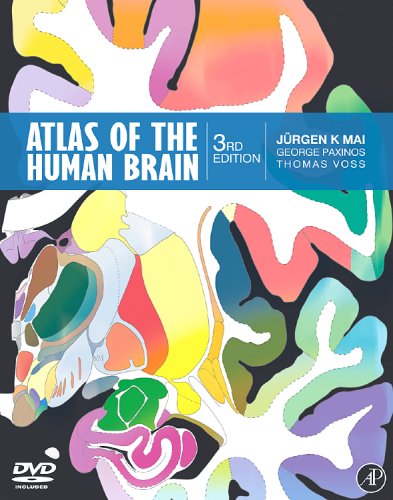
Atlas of the Human Brain, 3rd ed


High resolution human imaging requires a high resolution atlas. There is no atlas that approaches the accuracy and comprehensiveness of The Atlas of the Human Brain. This is the third, thoroughly revised, edition of the very successful atlas that received the Award of Excellence from the American Association of Publishers. Constructed by the recognized leaders in brain cartography, the atlas provides the most detailed and accurate delineations of brain structure available. It is intended as an atlas for day-to-day use in the research laboratory to identify macroscopic and microscopic structures in the human brain as collected in research data with a variety of methods. It will also be particularly useful for the interpretation of magnetic resonance imaging data. The Atlas includes a DVD with not only the atlas in electronic format, providing electronic versions of drawings and photographs to facilitate use of atlas contents with researchers' own material, but also 3D visualisation software which allows easy browsing of the images, and a feature to allow direct retrieval of brain areas using coordinates obtained in magnetic resonance imaging.

Download link: 

Anatomy for Diagnostic Imaging, 2nd ed.


This book covers the normal anatomy of the human body as seen in the entire gamut of medical imaging. It does so by an initial traditional anatomical description of each organ or system followed by the radiological anatomy of that part of the body using all the relevant imaging modalities. The third edition addresses the anatomy of new imaging techniques including three-dimensional CT, cardiac CT, and CT and MR angiography as well as the anatomy of therapeutic interventional radiological techniques guided by fluoroscopy, ultrasound, CT and MR. The text has been completely revised and over 140 new images, including some in colour, have been added. A series of 'imaging pearls' have been included with most sections to emphasise clinically and radiologically important points. The book is primarily aimed at those training in radiology and preparing for the FRCR examinations, but will be of use to all radiologists and radiographers both in training and in practice, and to medical students, physicians and surgeons and all who use imaging as a vital part of patient care. The third edition brings the basics of radiological anatomy to a new generation of radiologists in an ever-changing world of imaging.


Monday, April 4, 2011

Wednesday, March 9, 2011

B.D CHAURASIA


A wonderful book of anatomy by an Indian Author.


DOWNLOAD
OR
DOWNLOAD
password : www.anastomosis.co.nr

Neurology Board Review: An Illustrated Study Guide


Product Description:
Preparing the reader for the written portion of the Neurology Board examination and the Residency In-Service Training Examination (RITE) of the American Academy of Neurology, this reference provides a comprehensive review of the most critical topics in neurology. With the abundance of neuropathology and neuroimaging figures and neuroanatomy and neurophysiology illustrations, readers will not need to refer to multiple sources for a comprehensive review to prepare for the Neurology Boards and in-service examination.

PDF | 57 MB | no password

http://www.filefactory.com/file/d925e2/n/0411_neuroboardrev_rar
or
http://www.megaupload.com/?d=R7RCL6CK
or
DOWNLOAD

Grant's Dissector


Product Description:
Since 1940, when Dr. J.C. Boileau Grant created the first lab manual based on Grant's method of dissection, Grant's Dissector has clearly established its authority and preeminence as the "gold standard" of gross anatomy dissection manuals. In the last edition, the material was streamlined to focus on more accurate, specific and clear steps, based on market conditions and feedback. This edition continues to focus on the trend of reduced lab hours yet maintains the quality and reliability of Grant's original manual. Grant's Dissector, Fourteenth Edition features over 40 new figures to provide consistent appearance and include additional details, and is cross-referenced to the leading anatomy atlases, including Grant's, Netter's, Rohen, and Clemente.


DOWNLOAD

GRAYS ANATOMY 40TH EDITION


A universal landmark in medicine ever since Drs. Henry Gray and H.V. Carter published the first edition in 1858, Gray's Anatomy now celebrates its 150th anniversary! From state-of-the-art coverage of important new areas such as functional neuroimaging, embryogenesis, and biomechanics . . . through a comprehensively revamped, lavish full-color art program . . . as well as convenient access to the complete contents online, with downloadable illustrations, the new 40th Edition sets a new world standard for accuracy, clarity, and clinical relevance. It is the place to turn when you want to be sure about the anatomical considerations that pertain to safe and effective practice. You'll find it an invaluable clinical resource, a pleasure to consult, and a reference that you'll be proud to have on your shelf. 

Key Features 

* Fastidious editing by an esteemed group of clinicians and anatomists, with support from hundreds of contributors and specialist reviewers worldwide, ensures the most detailed and dependable coverage of anatomy available anywhere, including in-depth information on normal variants. 
* A regional organization collects all relevant material on each body area together in one place, for easy clinical reference. 

New to This Edition 

* Meticulously enhanced and updated artwork throughout - with 2,000 images now in full color - provide a more consistent, coherent view of every anatomical detail, providing practitioners across all specialties with the best, most clinically helpful visual understanding available. 
* 200 completely new radiologic, CT, MR, and other imaging studies vividly capture anatomy as it is primarily seen by physicians. 
* Significantly expanded clinical discussions emphasize considerations that may affect medical care. 
* Online access via Expert Consult allows you to reference the book's complete contents from any computer, fully searchable, and download thousands of detailed anatomical illustrations. 


DOWNLOAD

Sunday, March 28, 2010

Snell's Clinical Neuroanatomy, 6th edition (pdf)


Organized classically by system, this popular text gives medical and health professions students a complete, clinically oriented introduction to neuroanatomy. Each chapter begins with clear objectives, includes clinical cases, and ends with clinical notes, clinical problem-solving, and review questions. Hundreds of full-color illustrations, diagnostic images, and color photographs enhance the text. This Seventh Edition features new information relating the different parts of the skull to the brain areas, expanded coverage of brain development and neuroplasticity, and updated information on stem cell research.


LinkWithin

Related Posts Plugin for WordPress, Blogger...